Pf Joint Knee
Arthrocentesis of the knee joint aspiration.
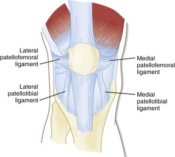
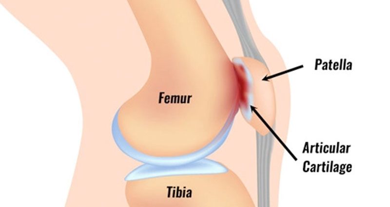
Pf joint knee. It can also occur as you get older. Joint space narrowing may occur from overuse of your joints. It is important to recognize that the retinacular support structure around the patella and the synovial lining in the joint are potential sources of pain as well as bone underlying defective cartilage of the pf joint. Patellofemoral pain syndrome may also result from overuse or overload of the pf joint.
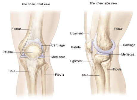
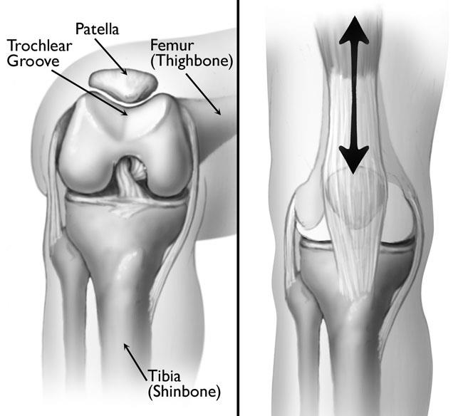
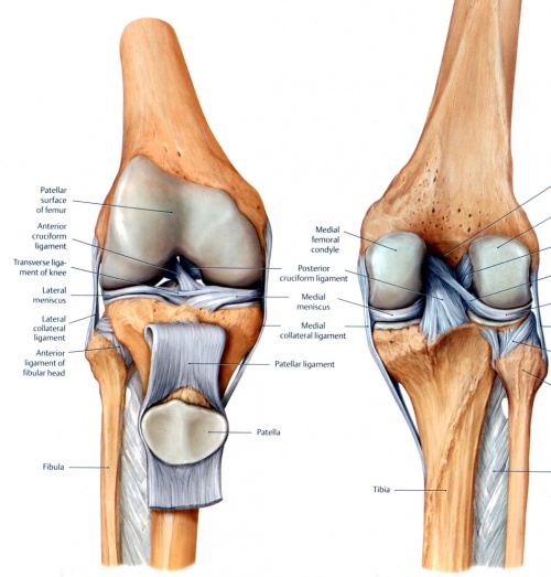
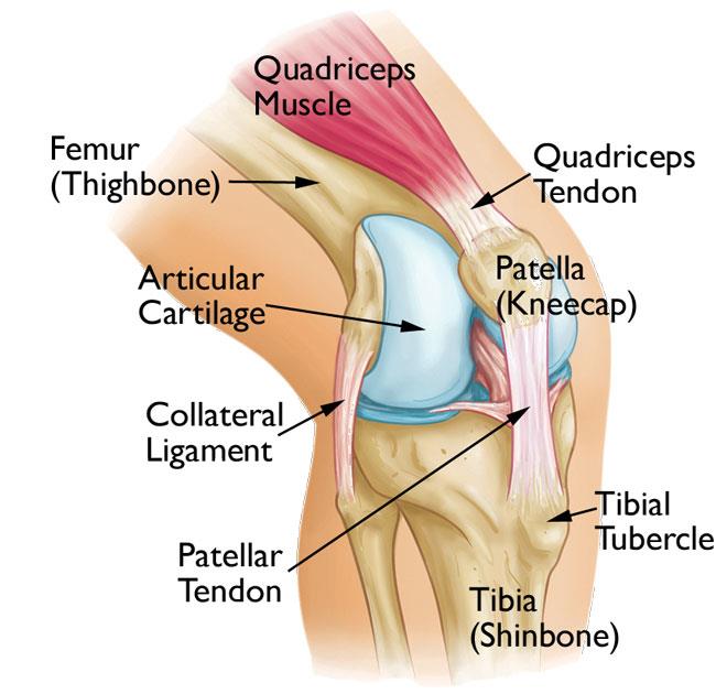
The patella has a configuration of a triangle with its apex directed inferiorly. In this article we shall examine the anatomy of the knee joint its articulating surfaces ligaments and neurovascular supply. A common cause of pf pain without instability is a lack of core muscle control and overuse of the knee. The knee joint is surrounded by a joint capsule with ligaments strapping the inside and outside of the joint collateral ligaments as well as crossing within the joint cruciate ligaments.
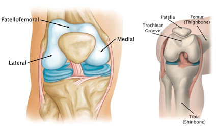
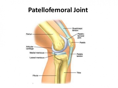
The patella protects the front of the knee joint. A needle is inserted into the joint space inside the knee and fluid is drawn out. Superiorly it articulates with the trochlea the distal articulating surface of the femur which are the main articulating surfaces of the patellofemoral joint. For this reason knee activity should be reduced until the pain is resolved.
The kneecap the patella joins the femur to form a third joint called the patellofemoral joint. Other risk factors such as obesity and muscle weakness can contribute to joint space narrowing. There is consistent but low quality evidence that exercise therapy for pfps reduces pain improves function and aids long term recovery. The patellofemoral joint is a unique and complex structure consisting of static elements bones and ligaments and dynamic elements neuromuscular system.
/Blausen_0597_KneeAnatomy_Side-5bbfb48fc9e77c0051f630c3.jpg)


